
What makes the heart beat? Your heart continues beating every moment of the day and night, even without you being aware of it. It may change its rate and rhythm for a few moments when you exercise or when you are excited, but it soon goes back to its regular pace without your conscious control. The cycle of contraction and relaxation of the heart muscle is generated by electrical impulses that are controlled by the conduction system of the heart, which may be influenced by factors such as temperature, exercise, and hormonal changes.
5 Elements of the Conduction Pathway
The conduction system of the heart controls its pumping action, which results in the delivery of blood to the different organs and tissues of the body. This conduction system is composed of a group of special cells found in the walls of the heart muscle, which send the electrical impulses and cause the heart muscle to contract.
It is made up of these elements:
The SA (Sinoatrial) node
- The SA node is found in the upper right chamber (atrium) of the heart.
- It initiates an impulse that causes depolarization and generates the action potential, an electrical event, which spreads out through the two upper chambers (atria) and to the AV node.
- It sets the pace of the beating of the heart.
The AV (Atrioventricular) node
- This group of cells is found between the atria and ventricles.
- It transmits electrical impulses from the atria, where the action potential is briefly delayed as they contract, to the AV bundle.
The AV bundle (Bundle of His)
- The electrical connection between the atria and the lower chambers of the heart (the ventricles) is through the AV bundle.
- It allows the movement of the action potential from the septum in the atria to the septum dividing the ventricles, and connects the AV node to the ventricular Bundle Branches.
The bundle branches
- These pass on the action potential to the heart’s interventricular septum.
Purkinje fibres
- These fibers begin at the interventricular septum down to the apex of your heart, and continue through the ventricular muscles (myocardium).
- They convey electrical impulses to the ventricles’ muscle cells.
- From an electrical event (action potential), a mechanical event (muscle contraction) occurs when the contractile cells act in a coordinated fashion, resulting in a heartbeat.
How Does the Conduction System Work?
The conduction system of the heart works this way:
Step 1: Pacemaker Impulse Generation
The SA node is known a natural pacemaker because it sets the pace of the heartbeat. It is where cardiac muscle contraction begins, from an impulse which causes the right and left atria to contract and push blood into the ventricles.
Step 2: AV Node Impulse Conduction
From the atria, the electrical signal spreads to the AV node, a group of cells found between the atria.
Step 3: AV Bundle Impulse Conduction
The electrical impulse then travels through the AV bundle of His, which divides into a right and left bundle branches.
Step 4: Purkinje Fibers Impulse Conduction
Then the action potential spreads through the Purkinje fibers, which causes the left and right ventricles to contract. The strong contraction of the ventricles causes the pumping of the blood from the right ventricle to the lungs, and from the left ventricle to the rest of the body. After the ventricles contract, they relax and get filled with more blood from the atria as an electrical impulse from the SA node begins the cardiac cycle again.
This video details how the electrical system of the heart work:
Cardiac Conduction System and Its Relationship with ECG
The action of the conduction system of the heart is recorded by anelectrocardiogram (ECG), which graphs the electrical impulses corresponding to the heartbeats. It is a useful tool to monitor the heart’s conduction system and may be used to detect abnormalities in function.
Heart and ECG Comparison
Ventricular contraction begins at the lower portion (apex) of your heart and moves upward and forces blood toward the large arteries, which are located above or superior to the ventricle.
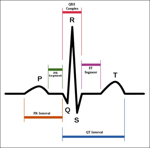
The heart’s electrical activity correlates with the ECG wave tracing:
- The P wave signals atrial depolarization, which corresponds to atrial contraction.
- The QRS complex signals ventricular depolarization, which corresponds to ventricular contraction.
- The T wave indicates ventricular repolarization, which corresponds to ventricular relaxation.
Problems That Can Go Wrong
Sometimes, the heart produces an irregular heartbeat or an arrhythmia. This may occur when:
- The sinus (SA) node changes its rate or rhythm.
- The conduction system of the heart is interrupted.
- Certain parts of the heart tissue take over the SA node and becomes another "pacemaker."
Arrhythmias cause heart palpitations and other symptoms such as dizziness, shortness of breath, or fainting.
You may need an ECG evaluation to monitor the rhythm of your heart. This painless test involves recording your heart’s electrical activity using small stickers that are attached to your chest. An abnormal electrical rhythm may or may not need treatment, which can include medications or surgery.
Watch this video to learn more about cardiac conduction system and its relationship with ECG.