
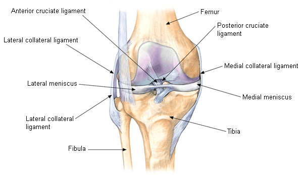
The medial collateral ligament is one of the main ligaments in the knee joint. It is found on the inner side of the knee. One end of the ligament is attached to the tibia while the other is attached to the femur. It prevents lateral movement of the tibia on the femur when valgus stress is put on the knee. Medial collateral ligament injury occurs during activities involving quick change of direction, twisting and bending, such as soccer or football. Injury can also occur when skiing or during other sports that have a lot of stop-and-go motion, weaving and jumping.
What Are the Symptoms of Medial Collateral Ligament Injury?
Types of Injury
Injuriesto the medial collateral ligament can be classified into 3 grades:
- The least severe is a grade 1 injury. This means that the ligament is stretched but not torn.
- A grade 2 injury means the ligament is partially torn, causing some instability in your knee joint.
- The most severe type is the grade 3 injury. This means your ligament is completely torn and you will experience knee instability.
Symptoms
Symptoms of a medial collateral ligament injury are similar to those of other knee problems, including:
- Catching or locking in the knee joint
- Feeling like your knee will give out when weight is placed on it
- Swelling of the knee joint
- Tenderness and pain along the inner parts of your knee
- When the injury occurs, you hear a popping sound
- Knee instability, which might be an indication of grade 2 or 3 injury
How Is Medial Collateral Ligament Injury Diagnosed?
The Valgus Stress Test
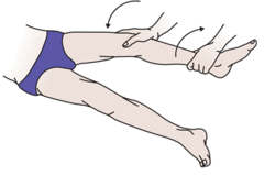
This test is done with the knee at 30 degrees of flexion and the hip abducted. Your leg is placed on the edge of a table, and the examiner places his/her thigh against yours for stability. Then, the examiner uses one hand to apply pressure on the anterior part of the ankle, and fingers of the other hand are placed exactly above the joint line to feel the amount of joint-line opening. The examiner can quantify the level of joint-line opening to be between 0 to 5mm, 5 to 10 mm or greater than 1 cm. This will tell if the injury is mild, moderate or severe.
Radiographic Assessment
An x-ray can be used to check for calcification at the beginning of the medial collateral ligament. An MRI can also be ordered to visualize the ligament using T2 weighted images. The examiner will able to see a concomitant meniscal tear.
How to Deal With Medial Collateral Ligament Injury?
The RICE Method
Try rest, ice, compression and elevation for the first 24-72 hours after sustaining the injury. Take a break from any movements or activities that cause pain to allow your knee time to heal. Use a cold therapy wrap or ice 10-15 minutes hourly at first, and reduce the frequency as the pain decreases. Do not put ice directly on your skin, use a wet towel or a cold therapy wrap. To protect the joint and reduce swelling, use knee support or compression bandages. Keep your injured knee elevated above your hip and allow gravity to drain out the fluid from the affected area.
Hinged Knee Brace
For grade 2 and 3 injuries, a hinged knee brace is recommended to limit knee movement and bending during healing.It has metal on the sides which helps prevent sideways motion.
Electrotherapy
During the ultrasound treatment, high frequency sound waves are applied to the injured area. A professional can use this method on you during the first serious stages of the injury to control pain and swelling. Tens or interferential involves application of electric currents to the area around the injury to assist with the swelling and pain.
Strapping
You can do this in the first stages of the injury as well as the later stages when you are going back to your routine. Good strapping techniques will offer more support compared to knee braces. However, the strap stretches after some time, so it needs to be re-applied to maintain effectiveness.
For a demonstration of how to do strapping of the medial collateral, watch this video:
Surgery
It is rare for a medial collateral ligament injury to require surgery. Surgery is necessary when the ligament is torn in such a way that it’s impossible for it to heal by itself, or when there are other ligament injuries in the picture. Arthroscopy, insertion of a tiny camera through small incisions made by the doctor, may be done before the surgery to determine the extent of your injury or whether there is presence of other injuries. If the ligament is torn where it is attached to the thighbone or shinbone, the surgeon can use a metal screw, bone staples, large stitches or anchor to reattach it. If the tear is in between the ligament, the surgeon will simply stitch it together.
Rehabilitation
A grade 1 injury will take 2-6 week to recover fully, while grade 2 and 3 will take 8-12 weeks.
- Exercise
Rehabilitation involves strengthening and mobility exercises. A variety of motion mobility exercises can be done to reinstate complete pain-free range of motion. Isometric strengthening exercises can be done in the first stages to prevent the muscle from wasting while the ligament heals and to maintain muscle strength. Exercises like leg presses, step-ups and mini squats can be helpful. However, you should stay away from exercise that requires sideways movements or change of direction. Wear a hinged knee brace during these exercises.
- Massage
During the early stages of the injury, massage should be avoided. As the ligament heals, light cross friction massage can be used, more so if there is still pain in the later stages of rehabilitation.