
Many people occasionally experiencepremature atrial complexes, a common type of heart arrhythmia. These irregular heartbeats occur in normal individuals and interrupt their normal sinus rhythm, which is heart rhythm that is generated by the heart structure called the sinus node. The sinus node is found at the top of the right upper chamber (atrium). It generates electrical signals that initiate heartbeats and controls your heart rate. A sinus rhythm normally generates 60-100 heartbeats per minute.
Premature atrial complexes or PACs consist of extra heartbeats that interrupt the sinus rhythm. They may occur anytime within a day in healthy individuals.
What Causes Premature Atrial Complexes?
PACs are early heartbeats that are caused by prematureelectrical impulses that arise
from anywhere within atrium. They are not generated by the sinus node. Although they occur in many healthy people, they may also be associated with a disease or heart injury.
In most cases, premature atrial complexes are not serious, but they may be significant if associated with atrial fibrillation. PACs are believed to trigger atrial fibrillation, an abnormal type of heart rhythm characterized by rapid, irregular beating that starts briefly and become longer, eventually persisting over time. There are usually no symptoms, but you may occasionally experience palpitations, shortness of breath, chest pain, orfainting. This condition may increase your risk of dementia, heart failure, and stroke. Ablation procedures are used to treat atrial fibrillation by eliminating PACs.
Electrocardiographic Features of Premature Atrial Complexes
A normal electrocardiogram (ECG) is a representation of the normal electrical activity of the heart muscle, which originates from the upper chambers (atria) and is conducted to the lower chambers (ventricles). The P wave represents the electrical signals from the atria. It is normally followed by a short delay, represented by a straight line, before it proceeds to the ventricles. Depolarization of the ventricles results in the largest part of the ECG signal, known as the QRS complex.Repolarization of the heart muscle (myocardium) is represented by the ST segment, an isoelectric line, and the T wave, which is seen as an upright deflection.
Premature atrial complexes have the following features:
- A non-sinus P wave that has a different shape compared to a sinus P wave.
- The abnormal P wave is followed by a QRS complex.
- A preceding T wave may hide the P wave, producing “peaked” waves or “camel humps” which may be mistaken for a premature junctional contraction.
- PACS that arise close to the atrioventricular node produce an inverted P wave.
- PACs reaching the SA node may cause it to “reset,” resulting in longer intervals before the next beats.
- PACs may be conducted abnormally, usually with the morphology of a right bundle branch block, which can be differentiated from premature ventricular contractions by thepresence of a P wave.
- PACs that arrive very early in the cardiac cycle may not trigger the ventricles so an abnormal P wave is not followed by a QRS complex, but is followed by a compensatory pause before the next beat.
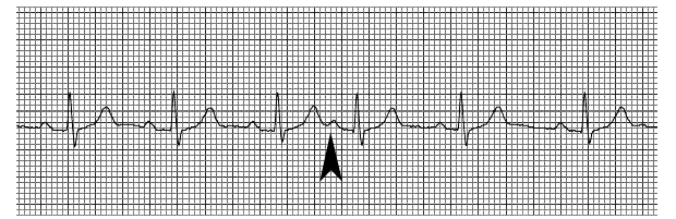
Single PACs
The example above shows an isolated PAC, which occurs after three normal heartbeats. The fourth beat is preceded by a P wave that occurs right after the T wave of the third beat. The QRS is normal in shape, indicating that this beat originated in the atrium.
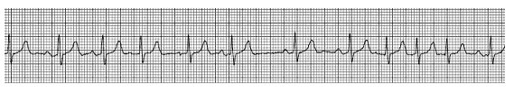
Pairs and Brief Runs of PACs
This example shows a normal sinus rhythm followed by a pair of PACs, then a normal beat. It is then followed by a single PAC, two normal beats, and a run of three PACs and a normal beat.
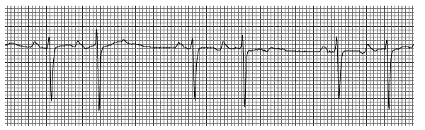
Bigeminal PACs
The example above shows bigeminal PACs, which are PACs that occur every other beat. When bigeminal PACs continue for more than 30 seconds, you may feel faintor lightheaded because your heart is not effectively pumping blood and your body is not getting enough oxygen.
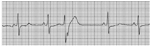
Aberrantly Conducted PACs
The example above shows an aberrant PAC (the third beat), which does not look like a normal beat. It looks different because the bundle branches that conduct the impulse from the atrium to the ventricle have not fully recovered from the previous beat since this beat occurred early. This caused the beat to slow down while traveling through the bundle branches, resulting in a change in its shape.
Premature Atrial Complexes Symptoms
Premature atrial complexes usually do not cause symptoms, although you may sometimes feel skipped beats, stronger heartbeats or a fluttering sensation in the chest.
Other associated symptoms that must prompt you to seek medical consultation include:
- lightheadedness orfainting
- becoming sweaty or pale
- difficulty breathing
- chest pain
- more than six episodes, with abnormal heartbeats coming in threes or more
- having more than 100 beats per minute at rest
How to Treat Premature Atrial Complexes
Premature atrial complexes are usually diagnosed with a Holter monitor, an electrocardiogram (EKG), or a cardiac event monitor.
Occasional PACs are common and do not usually indicate a health risk. Therefore, you will not need any treatment. Rarely, however, they may be associated with an underlying structural problem in the heart. PACs may trigger more serious arrhythmias such as atrial fibrillationor atrial flutter. An atrial flutter is an unstable heart rhythm originating in the atrium that causes very rapid heartbeats.
In these cases, the underlying condition must be treated. Medications like beta blockers may be prescribed for patients with symptomatic PACs.