The shoulder joint is one of the most important and freely moveable joints between the axial and the upper appendicular part of human body. Two main joints—the glenohumeral joint and the acromioclavicular joint in the shoulder help with the smooth motion for arms. The glenohumeral joint is responsible for most shoulder movements, yet for a further stability and to achieve optimal movement, the acromioclavicular joint needs to function normally. Usually, the shoulder joint refers to the glenohumeral joint. In this article, we will reveal the specific and detailed anatomy of the shoulder joint.
Shoulder Anatomy
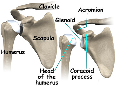
Bones: There are 3 bones in the shoulder—scapula, clavicle and the humerus. The 3 bones are joined together by ligaments, tendons and muscles, being covered by joint capsule so as to provide a platform for the arm to function and move.
Joints:The major joint of the shoulder is glenohumeral joint which consists of the ball shaped head of humerus and the socket shaped glenoid fossa of scapula.
Soft tissues: Bones and joints are covered with many soft tissues. Deltoid muscle can be the top layer of soft tissue under the skin. For the further layer under the deltoid muscle, it is the sub-deltoid bursa, which is fluid-filled water balloon like sac.
Besides, the shoulder also helps to articulate the scapula with the chest wall.
Shoulder Joint Anatomy
The main movement around the shoulder joint is to rotate the arm in a circular motion or to abduct out and away from the body, which is mainly assisted by the glenohumeral joint—also known as shoulder joint. Below we will reveal the shoulder joint anatomy.
 1. Bones
The humerus and scapula are two main bones forming the shoulder joint anatomy. The ball-like head of humerus gets loosely fit into the glenoid socket of the scapula, which is attributed mainly to the larger size of the head of humerus in comparison to the glenoid socket of scapula. This difference in the size does not allow the bones to fit perfectly without the aid of other soft tissues around the joint, which provide the joint with a stability factor.
2. Cartilage
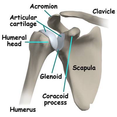
Labrum, the most important articular cartilage, lies between the humerus head and glenoid fossa of the shoulder joint. It is a ring shaped cartilage, thin at the center and thicker at the edges. The specificity of the labrum structure allows the large humerus head to fit into the smaller glenoid socket easily. Hence the labrum is not only important for joint stabilization, but also for smooth and free movement around shoulder joint by decreasing the friction to the least levels.
3. Muscles
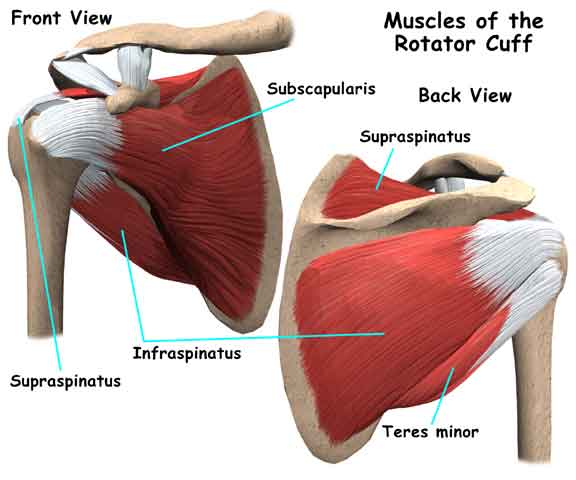 The main group of muscles in the shoulder joint is called rotator cuff, consisting of supraspinatus, subscapularis, infraspinatus and teres minor. The rotator cuff connects humerus to scapula in such a way, so as to fit the humeral head strong against the small glenoid socket of scapula.
The rotator cuff not only helps with the stabilization of the shoulder joint anatomy, but also helps to prevent the damage caused by the shoulder joint dislocation. Movements of the arm around the shoulder joint are also aided by the rotator cuff, particularly the rotatory movement of the arm. The supraspinatus muscle mainly helps with the movement of drawing the arm away from the body; hence it is more susceptible to injuries during flexion and abduction movements.
4. Ligaments
Ligaments around the shoulder joint further stabilize the joint anatomy and the movements around it. There are three glenohumeral ligaments and one coracoacromial ligament. The later joins the coracoids process of scapula to the humerus as the name suggests.
5. Joint Capsule and Bursae
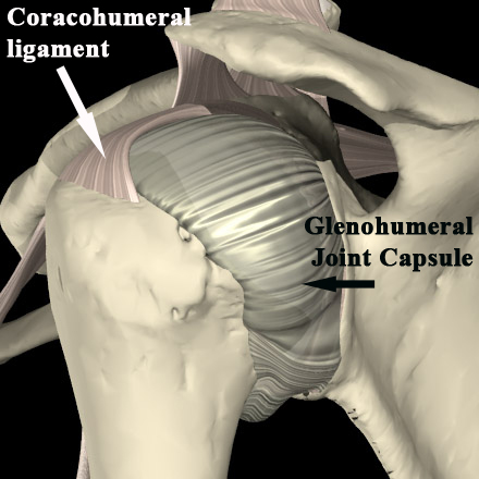 The joint capsule is a fibrous sheath the bounds and embeds the joint structures, which extends from the neck of humerus to the edges of glenoid cavity. The joint capsule is loosely attached so as to provide maximum movement around the shoulder joint. The friction is minimized by the synovial fluid which is released by synovial membrane adherent to the inner surface of the joint capsule. To further reduce the friction, the synovial fluid and bursae cushion the surface between the tendons of rotator cuff muscles and other joint structures. The most important bursae around the shoulder joint are subacromial and subscapular bursae. The subacromial bursae support the muscles around it, like deltoid and supraspinatus, while the subscapular bursae reduce the muscular damage around the shoulder joint during the joint mobility.
6. Nervous and Blood Supply
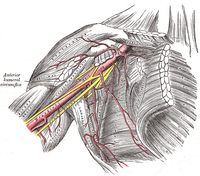 The blood supply of the shoulder joint is drawn from the anterior and posterior circumflex humeral arteries whose branches additionally form an anastomosis around the joint, making collateral supply function as well.
The nerve supply is derived from the roots C5 and C6 of the brachial plexus, including axillary nerve, suprascapular nerve and lateral pectoral nerves respectively.Thus, the Erb's palsy, the injury to C5 and C6 roots of the brachial plexus will affect the shoulder joint functions.
To better understand the shoulder joint anatomy, watch the video to have more details on visual information:
Common Issues on Shoulder
With the shoulder joint anatomy, you can better monitor common issues on shoulder. Below are some common ones:
- Frozen shoulder: In this condition, the movements of the shoulder joint are severely limiteddue to the inflammatory process going on in the joint anatomy. And you may experience pain and stiffness.
- Osteoarthritis: It is an outcome of aging along with routine wear and tear of the joint structures, but the shoulder joint is less commonly involved in osteoarthritis than the knee joint.
- Rheumatoid arthritis:It is an autoimmune condition where the body joints are attached by body's own defense soldiers, which can attack any joint in the body including the shoulder joint as well, causing pain and inflammation of the joint.
- Rotator cuff tear: Due to overuse, vigorous use or any injury around the shoulder region may lead to rotator cuff tear, which results in muscular pain and inflammation.
- Shoulder impingement: This condition refers to the pressing of the rotator cuff muscle against the acromion process of scapula when you lift your arms. Any presence of injury or inflammation will cause pain.
- Shoulder dislocation: The escape of the bones (usually the humerus) from the joint brim due to overhead extension mainly causes the joint to dislocate from its original place. In this condition there is moderate to severe pain in elevating the arm up, followed by a popping sensation around the joint.
- Shoulder tendonitis: This refers to the inflammation of any tendon which resides in the joint cavity of shoulder.
- Shoulder bursitis: It refers to the inflammation of the bursae that lies between the muscle tendons of the shoulder and other structures. Main symptoms include pain and excessive pressure which is more marked during movements.
- Labral tear: Labral tear usually occurs due to accidental injuries to the shoulder or its overuse. These tears mostly heal themselves without surgical interventions.
