
The spinal cord, which consists of the major nerve tract of vertebrates, runs down from the bottom of the brain through the passageway of the spinal column. This area is made up of all the nerve fibers that direct the reflex actions and convey the impulses that go back and forth to the brain.
When you look at the spinal cord cross section, it almost looks like a taffy candy that has a butterfly right in the middle. The gray matter that is in the central core makes up the dendrites and bodies of the neurons that make up the bundle. It is surrounded by the neuron axons or the white matter.
Spinal Cord Cross Section
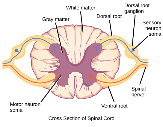
Looking at a cross section of the spinal cord, you would see gray matter shaped like a butterfly surrounded by white matter. The gray matter is the core and ends up to be four projections that are known as horns. At the back are two dorsal horns and away from the back are two ventral horns. The gray matter found here consists of interneurons, neurons, and glial cells, all part of the central nervous system. If you'd like to know more, click here for cross sections of spinal cord at eight different levels.
For clearer demonstration of the spinal cord cross section, watch the video below:
1. White Matter
The white matter has the nerve fibers that run up and down the length of the cord, they are called axons. This makes it possible for the different parts of the CNS communicate with each other. Every bundle of axons is a tract and it transmits specific information. The tracks that move upward are responsible for signals to the brain and the descending tracts send the signals from the brain to neurons throughout the body.
2. Gray Matter
In the center of the gray matter you will find the cerebrospinal fluid. The specific horns of the gray matter are responsible for different things. Each horn has different functions. The dorsal and ventral horns have the neurons that direct the skeletal muscles; the lateral horn has the cells that work the smooth muscle and supply cardiac.
3. More Spinal Cord Cross Section Facts to Know
- How Can You Tell Dorsal from Ventral?
The gray matter of the dorsal horns is thinner than the ventral horns.The dorsal roots can be cut in the cross section and come out from the horn area. The ventral roots look like axon streaks against the white matter.
- What Two Things Determine the Distribution of White Matter at a Particular Level of the Spinal Cord?
The amount of ganglion cells that are peripheral and send the axons upwards to the brain, and the amount of neurons that is in the brain projecting down to a specific spinal level.
Structure and Function of Spinal Cord
You may be surprised to find out that the spinal cord in an adult comes in at about 17 1/4 inches long, is about as wide as your thumb, and is just as thin as a straw that you drink from at the base.
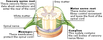
Protected by meninges, three layers of membrane just like those surrounding the brain; the bundle of nerves is thick and looks like jute rope that has hot dog casing around it. The nerve bundle has a cushion of cerebrospinal fluid between it and the meninges.
The end of the spinal cord is called the cauda equine because it looks like a horse’s tail with its cascade of nerves.
The Structure
Exiting through a big hole at the bottom of the skull, the spinal cord is covered by the vertebral column that protects it. The spinal nerves come out from the spaces between the bony arches in pairs. They are named for the area of the vertebral column from which they come. These areas include:
- Cervical or neck
- Thoracic or chest
- Lumbar or abdomen
- Sacral or pelvis
- Coccygeal or tailbone
Interesting enough, the spinal cord takes up around 2/3 of the vertebral canal where it rests. And that is because the vertebral column grows at a rate that is much faster than the spinal cord do,thus the cord will take up less and less room as the person grows. At this rate, the spinal cord will only extend up to the first lumbar vertebrae by the time a person reaches adulthood.
What Is the Spinal Cord Responsible For?
Basically the spinal cord is responsible for:
- The electrical communication between the various parts of the body and the brain. The spinal cord runs through several different levels of the trunkmaking it easier to communicate as the electrical signals are conducted through the cord.
- Walking, also referred to as locomotion. We take the simple act of walking for granted when it is really a series of commands sent to the leg groups telling them when to lift up and when to drop down. The signals go to the neurons who work the signals to the legs causing them to contract or extend. The movements continue back and forth repeatedly creating the act of walking.
- Reflexes are involuntary responses that go from the brain to the spinal cord to the nerves that make up the peripheral nervous system.
Here's is a video explaining clearly about the spinal cord: