
A normal functioning network of blood vessels is responsible for transporting oxygen and different nutrients to the brain. Two sets of vessels supply blood to the scalp, face and the brain, these are the left and the right vertebral arteries and the left and the right common carotid arteries. Carotid arteries have two divisions, the internal carotid artery supplies blood to the anterior three-fifths of the cerebrum (excluding the occipital and temporal lobes); whereas the external carotid artery supplies blood to the scalp and face. The vertebrobasilar arteries supply blood to the posterior two-fifths of the cerebrum present within the cerebellum, and the brainstem. 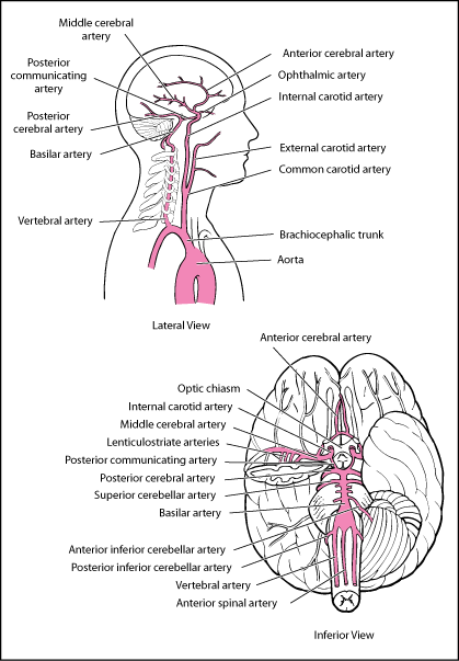
What Is the Posterior Cerebral Artery?
The carotid and vestibular arteries form a circle of communicated arteries at the base of the brain, this circle is termed as Circle of Willis. This circle gives rise to various other arteries such as the posterior cerebral artery (PCA), the middle cerebral artery (MCA) and the anterior cerebral artery (ACA). All these arteries extend to the brain covering the entire matrix. The vertebral arteries branch into posterior inferior cerebellar arteries (PICA). Due to the formation of a circle between vertebrobasilar and carotid arteries, if any main artery becomes obstructed; then arteries to which it supplies the blood (the distal smaller arteries) may receive blood from other arteries. This is called collateral circulation that serves as an alternate channel of circulation in order to prevent ischemia.
The basilar artery undergoes bifurcation at the site of midbrain, forming 2 posterior cerebral arteries. Both PCAs travel around the cerebral peduncles and branch into the midbrain forming a series of slender, long penetrating arteries that are responsible for supplying blood to the thalamus and hypothalamus. Hypothalamus is also partially surrounded by the Circle of Willis that helps supply extra blood to the hypothalamus by means of its perforators from other vessels.
Segments of PCA
- P1 segment: This segment extends from the point of terminationof the basilar artery and extends all the way up to posterior communicating artery (thereby spanning around the entire region of interpeduncular cistern).
- P2 segment: It rises from the posterior communicating artery, around the midbrain and is sectioned into two primary sub-segments; such as P2P posterior and P2A anterior. P2A is located within the crural cistern and is then joined to P2P in ambient cistern;hence it is also referred to as ambient segment in the medical literature.
- P3 segment: It is located within the quadrigeminal cistern, thus known as quadrigeminal segment.
- P4 segment: It is called cortical segment, including, for example, calcarine artery, that is present within the calcarine fissure.
Branches of PCA
- Lateral posterior choroidal arteries
- Medial posterior choroidal arteries
- Posterior communicating artery
- Perforators: Circumflex (short and long), Peduncular perforator (from P2), Thalamogeniculate perforator (this extends from P2), Posterior thalamoperforator (from P1) and Anterior thamaoperforator (from posterior communicating artery)
- Medial occipital artery: Parieto-occipital artery, Calcarine artery
- Lateral occipital artery: Posterior inferior temporal artery, Middle inferior temporal artery, Anterior inferior temporal artery
- Temporal branches: Posterior temporal artery, Anterior temporal artery
- Splenial artery
The Function of the Posterior Cerebral Artery
Posterior cerebral artery is responsible for supplying blood to the cerebellum, brain stem, inferior sections of temporal lobes and center of occipital lobes. This region includes calcarine cortex, often known as the primary visual cortex. The smaller branches of posterior cerebral artery transfer blood to midbrain, region of the optic path ways, thalamus and hippocampus.
PCA has central location in brain and it forms the lower portion of the Circle of Willis. This network of arteries also comprises of anterior and posterior communicating arteries as well as internal carotid, anterior cerebral, middle cerebral arteries. This fine network of arteries collectively transports the oxygenated blood to various parts of the brain.
The Posterior Cerebral Artery Stroke
Posterior cerebral artery stroke is rare compared to the stroke associated with the damage to the anterior circulation. Inadequate blood flow to any part of brain can lead to ischemic strokes. The metabolism of neurons can only tolerate a short period of inadequate glucose and oxygen supply, after which it develops irreversible damage. Cell death begins after approximately 6 minutes of inadequate blood supply. Ischemia is more likely to occur in large cortical neurons that are more metabolically active. Infarcts comprise of umbra or central area with high rate of cell death which is then surrounded by penumbra of tissues that contain stunned cells which may recover provided that the circulation is produced through collaterals or is reestablished.
Symptoms
Sustained PCA stroke may result in sensory and visual deficit. PCA stroke patients are less likely to experience any chronic disability unlike the patients of anterior cerebral, basilar, or middle cerebral infarctions.
Clinical signs and symptoms related to the occluded posterior cerebral artery are majorly dependent upon the site of occlusion. These symptoms may be:
- Hallucinations
- Verbal dyslexia
- Failure to see to-and-fro movements
- Color blindness
- Hemianopsia
- Contralateral hemiplegia
- Weber’s syndrome
- Thalamic perforate syndrome
- Thalamic syndrome
Affected Brain Functions
Stroke can affect the posterior cerebral artery as well as the occipital cortex and may cause alexia, which is characterized by inability to read. These strokes may also affect visual-spatial orientation, visual recognition and visual learning. Stroke affecting the posterior cerebral artery may also affect the brainstem or cerebellum resulting in slurred and slow speech. This condition is termed as dysarthria, which occurs as a consequence of damaged nerves that affect the muscles, responsible for controlling the jaw and tongue. Mostly the strokes are unilateral, i.e. they affect only the half part of the brain.