
Eye health is something that you need to become more aware of if you want to keep your vision sharp and avoid common eye diseases. However, there are many eye diseases that everyone is possible to experience when aging, like vitreomacular traction, which this article will talk about in detail.
What Is Vitreomacular Traction?
The retina is very important to our eyes. The macula, a part of retina, is the most sensitive tissue covering the inside back of the eye. Thus, macula is the key thing to do the specific visual 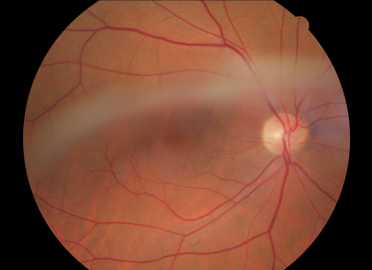 tasks. The eye itself is filled with a vitreous gel that serves to keep the retina flush to the macula. However, with age, the amount of gel in the eye decreases, causing the macula to detach from the retina. This results in the normal loss of fine focus. If the original adhesion between the macula and shrinking gel is too tight, then the disturbance to the central vision may be greater. People who experience this are said to have vitreomacular traction (VMT).
VMT is not common. It is estimated that it occurs in only 1 out of every 4400 persons. It is considered as a disease associated with aging. As the world's population gets older, the cases of VMT diagnosed are growing. This doesn't mean it has become more common, but that the age group it affects has grown larger.
What Are the Symptoms of Vitreomacular Traction?
Symptoms can be hard to detect in mild cases. As the vitreous membrane is pulled, it can stress the macula or tear it. This can cause blurriness and loss of central vision. Loss of side or peripheral vision is rare even in severe cases. The central vision loss is most accurately described as an increase in central vision blurriness and distortion that can interfere with daily activities.
How VMT affects a person is highly individual. Some people will only experience mild blurriness that can be corrected with glasses; while others will have such distorted central vision that they can no longer do the majority of their daily activities.
How to Diagnose Vitreomacular Traction
There are two tests used to diagnose vitreomacular traction. Both are done by retina specialists. The first is the OCT scan, or Optical Coherence Tomography scan. This is a simple retina scan that can be done with a little preparation. The second is a fluorescein angiogram. This is a photographic test that requires a dye be injected to make the vitreous membrane more easily seen on the film after. A series of photographs are taken to see how the dye passes through the back of the eye to diagnose the severity of the condition. The dye used for this test is well tolerated by most people and the test is painless.
How to Treat Vitreomacular Traction
The good news is that if you have only mild symptoms, no treatment is needed as the disruption to your vision is manageable with vision aids such as glasses. If you do have more severe symptoms, then your doctor may recommend surgery. During the surgery for vitreomacular traction, the vitreous membrane is separated from the eye and any underlying scar tissue is removed with micro-forceps. Your doctor may also prescribe a medication injection that will assist in this removal process. Most patients who have had this surgery done report that they can return to normal activities quickly, as well as being able to read up to two or three more lines on the eye chart compared to their sight ability before.